Median Rhomboid Glossitis Histology. Often the location can vary from center to the back of the tongue. Median rhomboid glossitis mrg also referred to as central papillary atrophy was once thought to be a developmental defect that occurred during embryogenesis caused by the failure of the tuberculum impar to be covered completely by the lateral processes of the tongue.
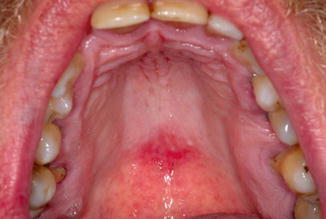
Often the location can vary from center to the back of the tongue. About 1 of the population gets affected by the median rhomboid glossitis. It is suggested that median rhomboid glossitis is the clinical expression of a localized chronic fungal infection with candida species.
It occurs in as many as 1 of adults.
The affected area of the tongue is missing its normal coating of finger like projections called filiform papilla which normally cover the entire top surface of the tongue. This paper reports a case of rhomboid glossitis in a 61 year old man who consulted for a painless raised lesion on the dorsum of the tongue in left paramedial not. Median rhomboid glossitis mrg also referred to as central papillary atrophy was once thought to be a developmental defect that occurred during embryogenesis caused by the failure of the tuberculum impar to be covered completely by the lateral processes of the tongue. Often the location can vary from center to the back of the tongue.
